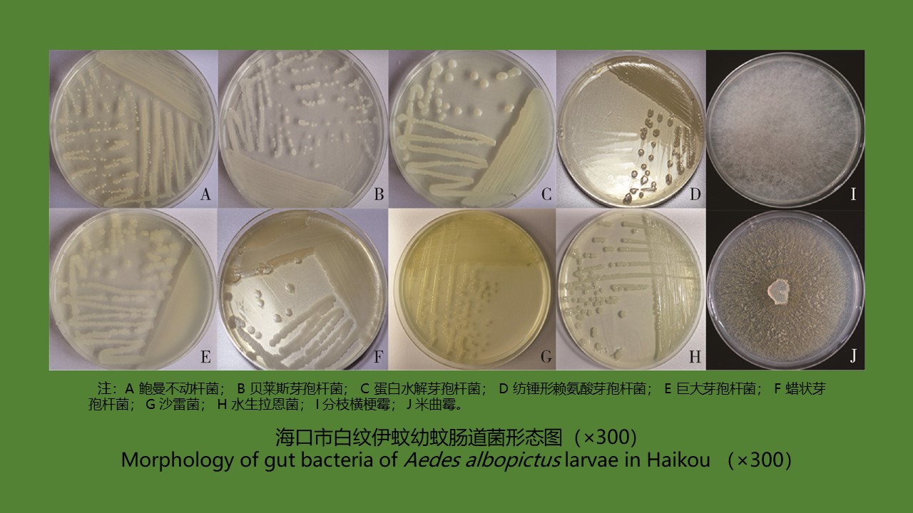
目的 通过对海口市及周边5个地点的白纹伊蚊幼蚊肠道微生物的分离与鉴定,研究白纹伊蚊肠道微生物菌群的种类和多样性。方法 2019年在海口市及周边5个地点采集白纹伊蚊幼蚊;对幼蚊肠道微生物进行PCR扩增,产物测序,通过美国国立生物技术信息中心(NCBI)数据库对幼蚊和肠道微生物序列进行核苷酸序列同源性比对分析。结果 白纹伊蚊幼蚊肠道微生物共鉴定出8株细菌,2株真菌,隶属于2门4科4属,优势菌群为厚壁菌门。海口市溪宜下村、大顶村,文昌市抱罗镇高堆村这3个地点的幼蚊肠道均分离出蜡状芽孢杆菌,仅临高县昌拱村的幼蚊中分离出真菌。海口市溪宜下村白纹伊蚊幼蚊肠道分离出的细菌种类最多,包括贝莱斯芽孢杆菌、纺锤形赖氨酸芽孢杆菌、蜡状芽孢杆菌和鲍曼不动杆菌。结论 海口市及周边5个地点白纹伊蚊幼蚊肠道细菌以厚壁菌门细菌为主;肠道微生物细菌丰富度高于真菌。
Objective To study the species and diversity of gut microbiota of Aedes albopictus larvae collected in Haikou and five surrounding sites, China. Methods Ae. albopictus larvae were collected in Haikou and five surrounding sites in 2019. Polymerase chain reaction amplification and product sequencing were performed for gut microbiota of the larvae. The nucleotide sequences of the larvae and gut microbiota were analyzed for homology through the National Center for Biotechnology Information database. Results A total of 8 strains of bacteria and 2 strains of fungi, belonging to 4 genera in 4 families of 2 phyla, were identified in the gut microbiota of Ae. albopictus larvae, with the dominant microflora from the Firmicutes. Bacillus cereus was isolated from the intestinal tract of the larvae in Xiyixia village and Dading village of Haikou city, and Gaodui village, Baoluo town of Wenchang city Fungi were isolated only from the larvae in Changgong village of Lingao county. Ae. albopictus larvae in Xiyixia village, Haikou had the most species of bacteria isolated from the intestinal tract, including B. velezensis, Lysinibacillus fusiformis, B. cereus, and Acinetobacter baumannii. Conclusion Gut bacteria of Ae. albopictus larvae are mainly from the Firmicutes in Haikou and five surrounding sites. The species diversity of bacteria is greater than that of fungi among gut microbiota.
[1] 贾德胜, 谭伟龙, 王长军, 等. 伊蚊传播疾病及其防治[J]. 中华卫生杀虫药械, 2017, 23(1):1-7. DOI:10.19821/j.1671-2781.2017.01.001. Jia DS, Tan WL, Wang CJ, et al. Aedes-borne diseases prevention and control[J]. Chin J Hyg Insect Equip, 2017, 23(1):1-7. DOI:10.19821/j.1671-2781.2017.01.001.(in Chinese)
[2] Scolari F, Casiraghi M, Bonizzoni M. Aedes spp. and their microbiota:A review[J]. Front Microbiol, 2019, 10:2036. DOI:10.3389/fmicb.2019.02036.
[3] Yadav KK, Bora A, Datta S, et al. Molecular characterization of midgut microbiota of Aedes albopictus and Ae. aegypti from Arunachal Pradesh, India[J]. Parasit Vectors, 2015, 8:641. DOI:10.1186/s13071-015-1252-0.
[4] Dong YM, Manfredini F, Dimopoulos G. Implication of the mosquito midgut microbiota in the defense against malaria parasites[J]. PLoS Pathog, 2009, 5(5):e1000423. DOI:10.1371/journal.ppat.1000423.
[5] Ricci I, Damiani C, Capone A, et al. Mosquito/microbiota interactions:From complex relationships to biotechnological perspectives[J]. Curr Opin Microbiol, 2012, 15(3):278-284. DOI:10.1016/j.mib.2012.03.004.
[6] Minard G, Mavingui P, Moro CV. Diversity and function of bacterial microbiota in the mosquito holobiont[J]. Parasit Vectors, 2013, 6(1):146. DOI:10.1186/1756-3305-6-146.
[7] Dennison NJ, Jupatanakul N, Dimopoulos G. The mosquito microbiota influences vector competence for human pathogens[J]. Curr Opin Insect Sci, 2014, 3:6-13. DOI:10.1016/j.cois.2014.07.004.
[8] Coon KL, Vogel KJ, Brown MR, et al. Mosquitoes rely on their gut microbiota for development[J]. Mol Ecol, 2014, 23(11):2727-2739. DOI:10.1111/mec.12771.
[9] Kim CH, Lampman RL, Muturi EJ. Bacterial communities and midgut microbiota associated with mosquito populations from waste tires in East-Central Illinois[J]. J Med Entomol, 2015, 52(1):63-75. DOI:10.1093/jme/tju011.
[10] 曾晓露. 海南地区疟疾疫情分布特征及环境影响因素研究[D]. 重庆:第三军医大学, 2015. Zeng XL. Study on the distribution characteristics of malaria epidemic situation and its environmental factors in Hainan province[D]. Chongqing:Third Military Medical University, 2015. (in Chinese)
[11] Li X, Tan XF, Chen QS, et al. Prodigiosin of Serratia marcescens ZPG19 alters the gut microbiota composition of Kunming mice[J]. Molecules, 2021, 26(8):2156. DOI:10.3390/molecules 26082156.
[12] Guégan M, Zouache K, Démichel C, et al. The mosquito holobiont:Fresh insight into mosquito-microbiota interactions[J]. Microbiome, 2018, 6(1):49. DOI:10.1186/s40168-018-0435-2.
[13] Chao J, Wistreich GA. Microbial isolations from the midgut of Culex tarsalis Coquillett[J]. J Insect Pathol, 1959, 1:311-318.
[14] Demaio J, Pumpuni CB, Kent M, et al. The midgut bacterial flora of wild Aedes triseriatus, Culex pipiens, and Psorophora columbiae mosquitoes[J]. Am J Trop Med Hyg, 1996, 54(2):219-223. DOI:10.4269/ajtmh.1996.54.219.
[15] Pidiyar VJ, Jangid K, Patole MS, et al. Studies on cultured and uncultured microbiota of wild Culex quinquefasciatus mosquito midgut based on 16S ribosomal RNA gene analysis[J]. Am J Trop Med Hyg, 2004, 70(6):597-603. DOI:10.1016/S0008-6215(97)00005-0.
[16] Rani A, Sharma A, Rajagopal R, et al. Bacterial diversity analysis of larvae and adult midgut microflora using culture-dependent and culture-independent methods in lab-reared and field-collected Anopheles stephensi-an Asian malarial vector[J]. BMC Microbiol, 2009, 9(1):96. DOI:10.1186/1471-2180-9-96.
[17] Boissière A, Tchioffo MT, Bachar D, et al. Midgut microbiota of the Malaria mosquito vector Anopheles gambiae and interactions with Plasmodium falciparum infection[J]. PLoS Pathog, 2012, 8(5):e1002742. DOI:10.1371/journal.ppat.1002742.
[18] Chandel K, Mendki MJ, Parikh RY, et al. Midgut microbial community of Culex quinquefasciatus mosquito populations from India[J]. PLoS One, 2013, 8(11):e80453. DOI:10.1371/journal.pone.0080453.
[19] Wu P, Sun P, Nie KX, et al. A gut commensal bacterium promotes mosquito permissiveness to arboviruses[J]. Cell Host Microbe, 2019, 25(1):101-112.e5. DOI:10.1016/j.chom.2018. 11.004.
[20] Campbell CL, Mummey DL, Schmidtmann ET, et al. Culture-independent analysis of midgut microbiota in the arbovirus vector Culicoides sonorensis (Diptera:Ceratopogonidae)[J]. J Med Entomol, 2004, 41(3):340-348. DOI:10.1603/0022-2585-41.3.340.

