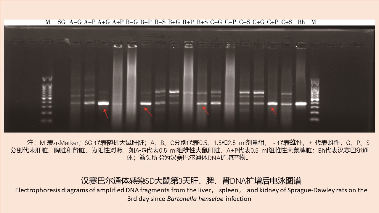
Objective To infect Sprague-Dawley (SD) rats with Bartonella henselae strains and measure the dynamic changes of bacteremia and antibody in rats. Methods SD rats were infected by subcutaneous multi-point injection of different concentrations of B. henselae suspension. Whole blood was collected and diluted for culture. The heart, liver, spleen, and kidney were homogenized and isolated for culture. The probable B. henselae colony nucleic acids were extracted, and the citrate synthase gene (gltA) was amplified by PCR for sequence analysis. The antibody changes were measured by the indirect immunofluorescence antibody assay. SPSS 19.0 software was used to analyze the differences in the positive rate between different tissues, different suspension concentrations, and different days of infection using the Chi-square test and two-way analysis of variance (ANOVA). Results A total of 90 whole blood samples, 82 tissue samples, and 21 serum samples were collected, all of which were negative for culture. For whole blood samples, the positive rate of the nucleic acid was 30.56% (22/72), and positive samples were found on days 1, 3, and 5, with the highest positive rate of 100% occurring in the low-dose group and high-dose group on day 3. For tissue samples, the positive rate of the nucleic acid was 22.86% (16/70), and positive samples were found on days 3 and 5; the renal tissues showed the highest positive rate (7/16) and the most persistent positive status (lasting 5 days), and the liver tissues had the second highest positive rate (5/18). The antibody was present in 6 of 18 serum samples detected, beginning on the 7th day since infection till the 14th day when the experiment ended. No significant difference was observed in the positive rate between different dose groups (F=3.243, P=0.082, by two-way ANOVA) or between different types of samples (χ2=7.655, P=0.057, by the Fisher's exact test). The positive rate showed a significant difference between different times of infection (F=11.770, P=0.001, by two-way ANOVA), and specifically between the 3rd day and the 1st, 5th, 7th, 10th, or 14th day (all P<0.05). Conclusion B. henselae can infect SD rats for a short time, with nucleic acid positive rate peaking on the 3rd day of infection. The antibody can last at least two weeks since infection.
[1] Oray M,Önal S,Akbay AK,et al. Diverse clinical signs of ocular involvement in cat scratch disease[J]. Turk J Ophthalmol,2017,47(1):9-17. DOI:10.4274/tjo.28009.
[2] Sykes JE,Henn JB,Kasten RW,et al. Bartonella henselae infection in splenectomized domestic cats previously infected with hemotropic Mycoplasma species[J]. Vet Immunol Immunopathol,2007,116(1/2):104-108. DOI:10.1016/j.vetimm.2006.12.004.
[3] 杨小冉,刘起勇,崔步云,等. BALB/c及昆明小鼠的汉赛巴尔通体实验室感染模型[J]. 中国媒介生物学及控制杂志,2008,19(1):41-43. DOI:10.3969/j.issn.1003-4692.2008.01.015. Yang XR,Liu QY,Cui BY,et al. Characterization of infective model in Bartonella henselae-specific immunity in BALB/c and KM mice[J]. Chin J Vector Biol Control,2008,19(1):41-43. DOI:10.3969/j.issn.1003-4692.2008.01.015.
[4] Arvand M,Ignatius R,Regnath T,et al. Bartonella henselae-specific cell-mediated immune responses display a predominantly Th1 phenotype in experimentally infected C57BL/6 mice[J]. Infect Immun,2001,68(10):6427-6433. DOI:10. 1128/IAI.69.10.6427-6433.2001.
[5] Polar RC,Orellana G,Caso WS,et al. Encephalitis with convulsive status in an immunocompetent pediatric patient caused by Bartonella henselae[J]. Asian Pacific J Trop Med,2016,9(6):610-613. DOI:10.1016/j.apjtm.2016.03.030.
[6] Chiaraviglio L,Duong S,Brown DA,et al. An immunocompromised murine model of chronic Bartonella infection[J]. Am J Pathol,2010,176(6):2753-2763. DOI:10.2353/ajpath.2010.090862.
[7] 白瑛,Kosoy MY,Maupin GO,等. 首次证实巴尔通体在我国云南鼠群中流行[J]. 中国人兽共患病杂志,2002,18(3):5-9. DOI:10.3969/j.issn.1002-2694.2002.03.001. Bai Y,Kosoy MY,Maupin GO,et al. Discovery of Bartonella species in rodents in Yunnan[J]. Chin J Zoonoses,2002,18(3):5-9. DOI:10.3969/j.issn.1002-2694.2002.03.001.
[8] 宋秀平,刘起勇,鲁亮,等. 海南省小型兽类巴尔通体的分离培养和序列分析[J]. 中国媒介生物学及控制杂志,2010,21(2):131-133. Song XP,Liu QY,Lu L,et al. Isolation and sequence analysis of Bartonella in small mammals in Hainan province[J]. Chin J Vector Biol Control,2010,21(2):131-133.
[9] 叶曦,姚美琳,李国伟. 福建省鼠形动物巴尔通体感染调查[J]. 中国人兽共患病学报,2006,22(8):779-781. DOI:10.3969/j.issn.1002-2694.2006.08.024. Ye X,Yao ML,Li GW. The investigation on the infection of Bartonella species in rodent hosts in Fujian[J]. Chin J Zoonoses,2006,22(8):779-781. DOI:10.3969/j.issn.1002-2694.2006. 08.024.
[10] 陈秀英,雷永良,柳付明,等. 浙江省丽水市鼠类巴尔通体的检测研究[J]. 中国卫生检验杂志,2013,23(1):118-119. Chen XY,Lei YL,Liu FM,et al. Study on the detection of Bartonella of Lishui rodents in Zhejiang province[J]. Chin J Heal Inspect,2013,23(1):118-119.
[11] 赖瑞青,徐晓红,黄洁玲,等. 33例猫抓病性淋巴结炎超声图像分析[J]. 广东医学院学报,2010,28(1):45-47. DOI:10.3969/j.issn.1005-4057.2010.01.019. Lai RQ,Xu XH,Huang JL,et al. Ultrasonographic analysis of 33 cases of cat scratch disease lymphadenitis[J]. J Guangdong Med Coll,2010,28(1):45-47. DOI:10.3969/j.issn.1005-4057.2010. 01.019.
[12] 宋秀平,栗冬梅,贾丽军,等. 内蒙古小型兽类巴尔通体感染情况调查[J]. 中国媒介生物学及控制杂志,2015,26(3):233-237. DOI:10.11853/j.issn.1003.4692.2015.03.004. Song XP,Li DM,Jia LJ,et al. Investigation of Bartonella infection in small mammals in Inner Mongolia,China[J]. Chin J Vector Biol Control,2015,26(3):233-237. DOI:10.11853/j.issn.1003.4692.2015.03.004.
[13] 左双燕. 我国黑龙江林区鼠型动物巴尔通体感染调查与分离鉴定[D]. 长沙:中南大学,2012. Zuo SY. Detection and isolation of Bartonella spp. in rodents in Helongjiang province of China[D]. Changsha:Central South University,2012.
[14] 王廷兆. 喜忧参半"宠物热"[J]. 环境,2002,5(10):27-28. Wang TZ. Pet fever[J]. Environment,2002,5(10):27-28.
[15] 刘纳新. 猫抓病23例明确诊断与治疗分析[J]. 中国误诊学杂志,2010,10(9):2204-2205. Liu NX. Diagnosis and treatment of cat scratch disease:an analysis of 23 cases[J]. Chin J Misd,2010,10(9):2204-2205.
[16] Brouqui PP,Houpikian XY,Lepedi H,et al. Virulence-related factors of Bartonella:observation of Bartonella infection in vitro and in vivo[M]//Kazar J,Toman R. Rickettsiae and Rickettsial Diseases Veda. Bratislava:Slovakia, 1996:622-627.
[17] Greene CE,Mcdermott M,Jameson PH,et al. Bartonella henselae infection in cats:evaluation during primary infection,treatment,and rechallenge infection[J]. J Clin Microbiol,1996,34(7):1682-1685. DOI:10.1128/jcm.34.7.1682-1685.1996.
[18] 栗冬梅,徐爱玲,宋秀平,等. 巴尔通体在自然感染的啮齿动物组织中的分布[J]. 中国媒介生物学及控制杂志,2021,32(2):181-187. DOI:10.11853/j.issn.1003.8280.2021.02.012. Li DM,Xu AL,Song XP, et al. Bartonella distribution in naturally infected rodent tissues[J]. Chin J Vector Biol Control,2021,32(2):181-187. DOI:10.11853/j.issn.1003.8280.2021.02.012.
[19] 康东梅,师灿南,开文龙,等. 宿主动物对不同宿主型猫栉首蚤吸血率和饱血率的影响[J]. 中国媒介生物学及控制杂志,2017,28(3):197-200. DOI:10.11853/j.issn.1003.8280.2017. 03.001. Kang DM,Shi CN,Kai WL, et al. Effect of hosts on blood-sucking and blood engorging in different host type cat fleas,Ctenocephalides felis[J]. Chin J Vector Biol Control,2017,28(3):197-200. DOI:10.11853/j.issn.1003.8280.2017.03.001.


