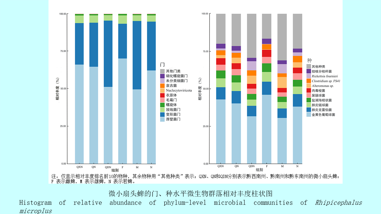
Objective To investigate the microbiota of the dominant tick species Rhipicephalus microplus in the minority autonomous prefectures of Guizhou province, China, and to provide a scientific basis for the prevention and control of tick-borne disease.Methods Rh. microplus ticks were collected from the body surface of cattle and sheep at 18 sampling sites located in Qiandongnan Miao and Dong Autonomous Prefecture, Qiannan Buyi and Miao Autonomous Prefecture, and Qianxinan Buyi and Miao Autonomous Prefecture in April and July of 2019 and 2020. The ticks were grouped by region, growth stage, and sex, with three parallel samples in each group, followed by metagenomic sequencing. The sequencing results were processed with quality control and assembly procedures before homology alignment with the non-redundant protein database of National Center for Biotechnology Information to obtain taxonomic annotation information. R (v. 3.6.3) and GraPhlAn (v. 1.1.3) were used for visual analysis, including species composition analysis, non-metric multidimensional scaling (NMDS) analysis, and analysis of similarities (ANOSIM).Results The dominant bacterial phylum for Rh. microplus was Firmicutes (60.70%), followed by Proteobacteria (33.76%) and Actinobacteria (4.53%). Staphylococcus aureus (38.29%) was the dominant bacterial species, followed by Klebsiella pneumoniae (7.79%) and Streptococcus pneumoniae (5.26%). Several tick-borne pathogens were detected, including various genotypes of spotted fever group Rickettsia and Anaplasma phagocytophilum (1.38%). R. fournieri (2.91%) was an emerging rickettsiae detected from ticks for the first time in China, and R. japonica (1.76%) and R. monacensis (0.04%) were common tick-borne Rickettsia genotypes in China. R. fournieri and R. japonica were mainly carried by male ticks. The NMDS analysis showed that the microbiota composition of Rh. microplus differed by growth stage and sex. The ANOSIM analysis indicated reasonable grouping, with greater intergroup differences than intragroup differences (R=0.147, P=0.014).Conclusions In the minority autonomous prefectures of Guizhou province, Rh. microplus ticks have a diverse microbiota composition, carrying pathogens that may cause spotted fever and human granulocytic anaplasmosis. Therefore, vector surveillance and pathogen detection should be strengthened, and effective control measures should be taken to reduce the risk of tick-borne diseases.
[1] Wikel SK. Ticks and tick-borne infections:Complex ecology,agents,and host interactions[J]. Vet Sci,2018,5(2):60. DOI:10.3390/vetsci5020060.
[2] Xiang LL,Poźniak B,Cheng TY. Bacteriological analysis of saliva from partially or fully engorged female adult Rhipicephalus microplus by next-generation sequencing[J]. Antonie Van Leeuwenhoek,2017,110(1):105-113. DOI:10.1007/s10482-016-0780-8.
[3] Gómez GF,Isaza JP,Segura JA,et al. Metatranscriptomic virome assessment of Rhipicephalus microplus from Colombia[J]. Ticks Tick-borne Dis,2020,11(5):101426. DOI:10.1016/j.ttbdis. 2020.101426.
[4] Segura JA,Isaza JP,Botero LE,et al. Assessment of bacterial diversity of Rhipicephalus microplus ticks from two livestock agroecosystems in Antioquia,Colombia[J]. PLoS One,2020,15(7):e0234005. DOI:10.1371/journal.pone.0234005.
[5] Anderson JF,Magnarelli LA. Biology of ticks[J]. Infect Dis Clin North Am,2008,22(2):195-215. DOI:10.1016/j.idc.2007. 12.006.
[6] Galay RL,Llaneta CR,Monreal MKFB,et al. Molecular prevalence of Anaplasma marginale and Ehrlichia in domestic large ruminants and Rhipicephalus (Boophilus) microplus ticks from Southern Luzon,Philippines[J]. Front Vet Sci,2021,8. DOI:10.3389/fvets.2021.746705.
[7] 杨茂生,吴位珩,杨莉,等. 贵州微小牛蜱的检测与防制研究[J]. 贵州农业科学,2009,37(11):157-159. DOI:10.3969/j.issn.1001-3601.2009.11.049.Yang MS,Wu WH,Yang L,et al. Study on detection and control of Boophilus microplus in Guizhou[J]. Guizhou Agric Sci,2009,37(11):157-159. DOI:10.3969/j.issn.1001-3601.2009.11.049.(in Chinese)
[8] Maldonado-Ruiz LP,Neupane S,Park Y,et al. The bacterial community of the lone star tick (Amblyomma americanum)[J]. Parasit Vector,2021,14(1):49. DOI:10.1186/s13071-020-04550-z.
[9] 杨小娜,张琳,侯学霞,等. 16S rDNA全长高通量测序在蜱媒病原生物多样性研究中的应用[J]. 中国媒介生物学及控制杂志,2021,32(4):404-411. DOI:10.11853/j.issn.1003.8280. 2021.04.004.Yang XN,Zhang L,Hou XX,et al. Application of 16S rDNA full-length high-throughput sequencing in the study of tick-borne pathogen biodiversity[J]. Chin J Vector Biol Control,2021,32(4):404-411. DOI:10.11853/j.issn.1003.8280.2021.04.004.(in Chinese)
[10] 黄邵军,张一,罗学辉,等. 基于高通量测序技术分析浙江省余姚市蜱中携带细菌菌群多样性[J]. 疾病监测,2020,35(7):642-645. DOI:10.3784/j.issn.1003-9961.2020.07.019.Huang SJ,Zhang Y,Luo XH,et al. High-throughput sequencing based analysis on diversity of pathogens carried by ticks in Yuyao,Zhejiang[J]. Dis Surveill,2020,35(7):642-645. DOI:10.3784/j.issn.1003-9961.2020.07.019.(in Chinese)
[11] 向昱龙,周敬祝,刘英,等. 贵州省部分地区蜱及其携带细菌调查[J]. 中国媒介生物学及控制杂志,2022,33(1):148-152. DOI:10.11853/j.issn.1003.8280.2022.01.027.Xiang YL,Zhou JZ,Liu Y,et al. An investigation of ticks and tick-borne bacteria in some areas of Guizhou province,China[J]. Chin J Vector Biol Control,2022,33(1):148-152. DOI:10.11853/j.issn.1003.8280.2022.01.027.(in Chinese)
[12] Matei IA,Estrada-Peña A,Cutler SJ,et al. A review on the eco-epidemiology and clinical management of human granulocytic anaplasmosis and its agent in Europe[J]. Parasite Vector,2019,12(1):599. DOI:10.1186/s13071-019-3852-6.
[13] Diop A,Barker SC,Eberhard M,et al. Rickettsia fournieri sp. nov.,a novel spotted fever group rickettsia from Argas lagenoplastis ticks in Australia[J]. Int J Syst Evol Microbiol,2018,68(12):3781-3784. DOI:10.1099/ijsem.0.003057.
[14] Li H,Li XM,Du J,et al. Candidatus Rickettsia xinyangensis as cause of spotted fever group rickettsiosis,Xinyang,China,2015[J]. Emerg Infect Dis,2020,26(5):985-988. DOI:10.3201/eid2605.170294.
[15] 韩婧,贺真,邵中军. 常见蜱传立克次体的研究进展[J]. 中华卫生杀虫药械,2022,28(1):86-89. DOI:10.19821/j.1671-2781.2022.01.024.Han J,He Z,Shao ZJ. The research progress of common tick-borne rickettsia[J]. Chin J Hyg Insect Equip,2022,28(1):86-89. DOI:10.19821/j.1671-2781.2022.01.024.(in Chinese)
[16] Li JB,Hu W,Wu T,et al. Japanese spotted fever in Eastern China,2013[J]. Emerg Infect Dis,2018,24(11):2107-2109. DOI:10.3201/eid2411.170264.
[17] Qin XR,Han HJ,Han FJ,et al. Rickettsia japonica and novel Rickettsia species in ticks,China[J]. Emerg Infect Dis,2019,25(5):992-995. DOI:10.3201/eid2505.171745.
[18] Narra HP,Sahni A,Alsing J,et al. Comparative transcriptomic analysis of Rickettsia conorii during in vitro infection of human and tick host cells[J]. BMC Genomics,2020,21(1):665. DOI:10.1186/s12864-020-07077-w.
[19] Sentausa E,El Karkouri K,Robert C,et al. Sequence and annotation of Rickettsia sibirica sibirica Genome[J]. J Bacteriol,2012,194(9):2377. DOI:10.1128/JB.00150-12.
[20] Li H,Fu XY,Jiang JF,et al. Severe illness caused by Rickettsia sibirica subspecies sibirica BJ-90 infection,China[J]. Emerg Microbes Infect,2017,6(1):1-3. DOI:10.1038/emi.2017.95.
[21] Jia N,Jiang JF,Huo QB,et al. Rickettsia sibirica subspecies sibirica BJ-90 as a cause of human disease[J]. N Engl J Med,2013,369(12):1176-1178. DOI:10.1056/NEJMc1303625.
[22] Ye XD,Sun Y,Ju WD,et al. Vector competence of the tick Ixodes sinensis (Acari:Ixodidae) for Rickettsia monacensis[J]. Parasite Vector,2014,7:512. DOI:10.1186/s13071-014-0512-8.
[23] Li W,Liu L,Jiang X,et al. Molecular identification of spotted fever group rickettsiae in ticks collected in central China[J]. Clin Microbiol Infect,2009,15:279-280. DOI:10.1111/j.1469-0691.2008.02235.x.
[24] 周琦,贺真,邵中军. 嗜吞噬细胞无形体的流行特征及临床诊断进展[J]. 中华卫生杀虫药械,2022,28(2):184-187. DOI:10.19821/j.1671-2781.2022.02.022.Zhou Q,He Z,Shao ZJ. Epidemiological characteristics and progress in clinical diagnosis of Anaplasma phagocytophilum[J]. Chin J Hyg Insect Equip,2022,28(2):184-187. DOI:10.19821/j.1671-2781.2022.02.022.(in Chinese)
[25] 刘增加,郑龙,张爱勤,等. 人粒细胞无形体病临床流行病学与防治研究现状[J]. 中华卫生杀虫药械,2018,24(5):417-422. DOI:10.19821/j.1671-2781.2018.05.001.Liu ZJ,Zheng L,Zhang AQ,et al. Research progress of clinical epidemiology,prevention and treatment on human granulocytic anaplasmosis[J]. Chin J Hyg Insect Equip,2018,24(5):417-422. DOI:10.19821/j.1671-2781.2018.05.001.(in Chinese)
[26] Wu XB,Na RH,Wei SS,et al. Distribution of tick-borne diseases in China[J]. Parasit Vectors,2013,6:119. DOI:10.1186/1756-3305-6-119.
