Chinese Journal of Vector Biology and Control ›› 2023, Vol. 34 ›› Issue (6): 723-727.DOI: 10.11853/j.issn.1003.8280.2023.06.003

• Experimental Study • Previous Articles     Next Articles

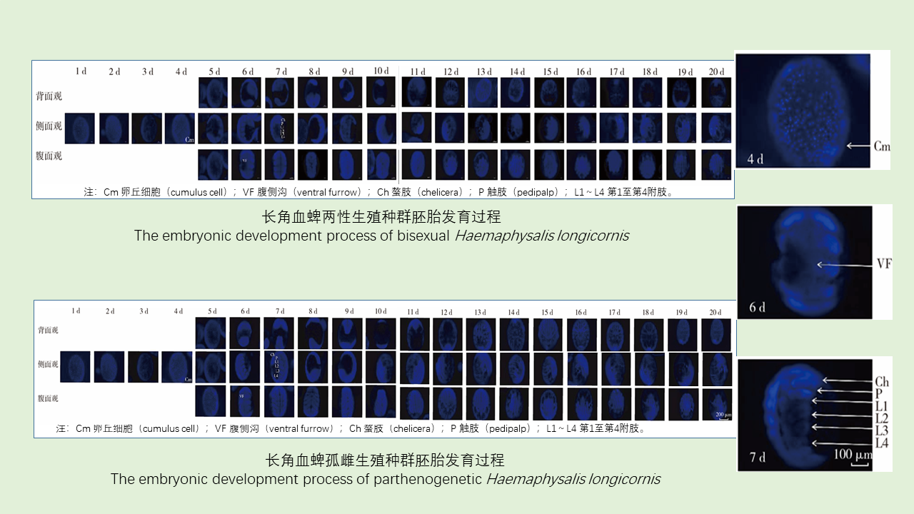
The embryonic development process of Haemaphysalis longicornis under laboratory conditions

CHEN Kai-li1, YANG Chen1, ZHANG Yu-fan1, LI Si-si1,2, LIU Jing-ze1, ZHANG Yan-kai1   

  1. 1. Hebei Key Laboratory of Animal Physiology, Biochemistry and Molecular Biology, College of Life Sciences, Hebei Normal University, Shijiazhuang, Hebei 050024, China;
    2. College of Life Science, Hengshui University, Hengshui, Hebei 053000, China
  • Received:2023-04-12 Online:2023-12-20 Published:2023-12-26
  • Supported by:
    National Natural Science Foundation of China (No. 31802008); Natural Science Foundation of Hebei Province of China (No. C2022205019)

实验室条件下长角血蜱胚胎发育过程

陈凯丽1, 杨晨1, 张雨凡1, 李思思1,2, 刘敬泽1, 张艳凯1   

  1. 1. 河北省动物生理生化重点实验室, 河北师范大学生命科学学院, 河北 石家庄 050024;
    2. 衡水学院生命科学学院, 河北 衡水 053000
  • 通讯作者: 张艳凯,E-mail:zyk412326@163.com
  • 作者简介:陈凯丽,女,在读硕士,主要从事蜱类生理生态学研究,E-mail:15830222006@163.com
  • 基金资助:
    国家自然科学基金(31802008);河北省自然科学基金(C2022205019)

Abstract: Objective To study the embryonic development of bisexual and parthenogenetic Haemaphysalis longicornis, so as to provide a basis for dynamic prediction and control of this species.Methods The embryonic development process of H. longicornis was recorded after embryo fixation and 4',6-diamidino-2-phenylindole staining under laboratory conditions (temperature, [26±1] ℃; relative humidity, [75±5]%; photoperiod, 16L∶8D). The independent samples t-test was used for comparison of differences between groups.Results The embryonic developmental period of parthenogenetic ticks ([24.6±1.7] d) was significantly longer than that of bisexual ticks ([22.5±3.5] d) (t=2.699, P=0.010). There was no significant difference in embryonic development between the two reproductive populations. The embryonic development process was generally as following: on day 1, the nucleus could be observed in the early embryo; on day 4, the number of cells increased significantly, and the blastoderm appeared; on day 6, the embryonic body segments and ventral furrow appeared; on day 7, the head, thorax, and growth zone could be clearly distinguished, the anlagen of the three pairs of legs (1L-3L) became clear, and the fourth leg pair (4L) was significantly shorter than the first three leg pairs; on day 12, the ventral furrow closed, the fourth pair of legs began to degenerate, the other three pairs of legs became larger, the embryo contracted to the anterior ventral region, and the dorsal region began to close; on day 16, the dorsal region continued to close, and the position of the chelicerae on the head changed; on day 20, dorsal closure was completed, and the embryo began to hatch.Conclusion This study presented the embryonic developmental process of H. longicornis under laboratory conditions, providing a reference for dynamic prediction and control of H. longicornis populations.

Key words: Haemaphysalis longicornis, Embryonic development, Embryo fixation, 4',6-Diamidino-2-phenylindole staining, Morphological change

摘要: 目的 在实验室条件下观察两性和孤雌生殖方式长角血蜱的胚胎发育过程,为该种群数量动态预测和控制提供依据。方法 在温度为(26±1) ℃,相对湿度为(75±5)%,光周期(L∶D)为16 h∶8 h条件下,应用胚胎固定和4',6-二脒基-2-苯基吲哚(DAPI)染色技术对长角血蜱胚胎发育过程进行观察描述。组间差异比较采用独立样本t检验。结果 孤雌生殖长角血蜱胚胎发育历期[(24.6±1.7) d]显著长于两性生殖长角血蜱[(22.5±3.5) d](t=2.699,P=0.010)。两种生殖种群胚胎发育过程无明显差异。胚胎发育过程大致分为:发育1 d,初期胚胎能够识别到细胞核;发育4 d,细胞数量显著增加,胚盘出现;发育6 d,胚胎体节和腹侧沟出现;发育7 d,能够明显区分出头部、胸部、生长区,3对附肢(1L~3L)原基清晰,第4附肢(4L)明显短于前3对附肢,发育12 d,腹侧沟关闭,第4附肢开始退化,另外3对附肢变大,胚胎收缩到前腹区,背侧开始闭合;发育16 d,背侧闭合继续发生,头部的螯肢定位发生改变;发育20 d,背侧闭合完成,胚胎开始孵化。结论 明确了长角血蜱胚胎在实验室条件下的发育过程,为该蜱数量动态的预测和控制提供了依据。

关键词: 长角血蜱, 胚胎发育, 胚胎固定, 4', 6-二脒基-2-苯基吲哚染色, 形态变化

CLC Number: